Polarisation imaging
Polarimetry is the study and measurement of the polarisation state of light and is a popular and useful tool in science today. Applications vary from astronomy, microscopy and biomedical diagnosis to more fundamental crystallographic, material and single molecule studies. Whilst the polarisation state of light itself, can be used to transmit information, hence presenting new possibilities in communications and optical data storage, changes in polarisation induced by an object can, alternatively, be used to expose new material or sample properties to scientific scrutiny, such as material birefringence, chirality or molecular orientation. Our research in polarimetry includes the following:
Polarisation microscopy

Incorporating polarisation sensitive elements into existing optical imaging systems enables polarisation imaging, in which either the polarisation state of the incident light is mapped (Stokes imaging) or the polarisation properties of the object are considered (Mueller matrix imaging). High resolution imaging, however requires high numerical aperture lenses, which further transform the polarisation. Development of accurate theoretical models and experimental calibration procedures, is thus important for accurate quantitative polarisation measurements. Recent decades have also seen a number of single molecule imaging techniques, such as fluorescence microscopy, STED and other localisation imaging modalities. Such techniques typically only measure the optical intensity from a molecule and eschew the information carried by the polarisation of light. We, however, consider how polarisation measurements can be used to yield additional information, for example for determining the 3D orientation of single fluorescent molecules, important in, for instance, studies of cell dynamics and monitoring their local environmental conditions.
Optimisation of polarimeters

In biological applications light levels are often kept relatively low so as to avoid sample damage, whereas in quantum or astronomically applications photon numbers are frequently inherently low. Consequently, the presence of noise, be it fundamental shot noise or technical noise from scattering, can be quite detrimental to measurement accuracy. In polarimetry, this problem is further compounded by the need to make multiple measurements of different polarisation states. Optimising the design of polarimeters is thus an important component of high accuracy studies. Using elements of discrete computational geometry, information and statistical estimation theory we consider how improvements can be made through use of a priori knowledge of the system, choice of different measurement bases and enforcing physical constraints.
Spectropolarimetry

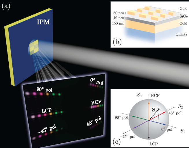
In addition to the additional information afforded by polarisation measurements, spectral degrees of freedom can yield details of the chemical composition of a sample, particle velocity, local magnetic and electric fields and more. Spectral-polarimetry therefore constitutes an exhaustive analytic optical tool. Spectropolarimetry can be practically realized using a number of experimental architectures, which differ through the means of spectral and polarization discrimination. Separation of individual spectral components is typically achieved by means of either a tunable optical or a dispersive element, such as a prism or grating structure. On the other hand, differing polarization components, are typically found using sequential measurements with variable polarisation elements or simultaneously using a division of amplitude polarimeter or polarization gratings. We have recently demonstrated a novel architecture employing plasmonic metasurfaces (nano-structured thin metallic films) enabling simultaneous determination of both spectral and polarisation information using a compact device, promising many applications in polarization imaging, quantum communication and quantitative sensing. Capitalising on our work on Müller matrix microscopy, we are also developing a spectroscopic version of this microscope that will permit 3D functional imaging of plasmonic structures and photonic crystals.
Microresonators
Optical micro-resonators are micron sized structures which allow light to propagate in a closed path and have in turn enabled a wealth of fundamental physical, chemical and biological studies, as well a host of technological applications. Confining light to micrometer scale resonators for example can be used for sensing, as a nonlinear light source, or as an optical element in photonic circuits.
Theory and simulation of resonant modes

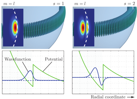
Resonant phenomena in optical cavities are frequently dependent on the precise geometric properties, such as size, shape, and composition, of the supporting structure. Moreover, the wave nature of light implies that when light is confined in a dielectric microstructure and brought to interfere with itself, only specific optical frequencies can be supported and reside within the cavity without suffering large losses. Knowledge of the mode structure and associated electromagnetic fields underpins the technological exploitation of microresonators. Whispering gallery modes, which are surface type modes supported in closed concave resonators, such as micro-spheres or toroids, are particulary promising due to their ultra high quality factors and sensitivity to the local environment, and thus form the focus of our efforts in this area. Specifically, we develop both approximate analytic and numerical models for the simulation of whispering gallery modes and how they are perturbed by external factors, such as particle binding, temperature changes, solute diffusion and application of external fields. We also consider the properties and behaviour of optically anisotropic and inhomogeneous resonators, which offers interesting applications in selectively coupling to microresonators, or high sensitivity monitoring of diffusion kinetics and degradation of polymers.
WGM sensors
Resonant microcavities provide a powerful platform on which the next generation of high-performance optical sensors can be realised. If the microcavity geometry or material properties change, e.g. due to temperature variations, a change in resonance parameters can be detected, for instance, by monitoring changes in light intensity or frequency shifts. Optical resonators can be used for a wide assortment of detection tasks; for instance, functionalised microcavities can detect specific biomolecules, whereas microcavities modified with magnetorestrictive materials can find use as magnetometers.

One specific class of resonant optical sensors that we consider are those based on microcavities supporting whispering gallery modes (WGMs) which offer an extreme level of sensitivity. A longstanding goal for biomedical detectors, environmental monitors, and biosensors in the life sciences has been to detect single molecules and their interactions. In the drive for ever more sensitive detection the need for system optimisation and enhancement is thus paramount. We pursue an array of strategies to achieve this goal. One line of our work has been to investigate how whispering gallery mode resonators can be enhanced through use of plasmonic nanoparticles so as to facilitate the detection of single DNA strands, in addition to monitoring their hybridisation dynamics. We also employ ideas from information and statistical estimation to optimise sensor parameters. and have furthermore considered alternative resonator geometries, such as droplet sensors.
Quantification of biomolecular properties is also an important and sought after capability since it can aid in particle discrimination and hence disease diagnosis, provide insight into protein function and facilitate studies of reaction kinetics or conformational changes. Through use of temperature stabilised resonators, multi-mode or polarimetric measurements we have been investigating how thermal properties, protein size and geometric parameters can also be extracted on a WGM sensing platform.
Nanoplasmonics
Plasmonics is the study of the coupling between photons and electron oscillations in conducting materials, such as metals, and has seen tremendous advancement in the last few decades due to improvements in nanofabrication techniques. Structures supporting plasmons can give strong localised fields, guiding of light and a means to control light-matter interactions and thus ‘plasmonics’ research can span a broad range of topics. Research in the group considers a variety of aspects of plasmonics as outlined below.
Surface plasmon resonance (SPR) sensors
Surface plasmons polaritons (SPPs) are electromagnetic excitations, coupled to coherent oscillations of electrons, propagating along the interface between, typically, a metal and a dielectric. Perpendicular to the interface SPPs experience an exponential decay in their amplitude and are hence quasi-2D bound modes. Evanescent wave contributions can dominate in the near field and can give rise to large local intensities and endow SPPs with a high sensitivity to perturbations in the vicinity of the surface. Consequently SPPs have found myriad uses, such as bulk sensing, high resolution spectroscopy (e.g. SERS) and microscopy.
Mesoscopic plasmonics

As with all wave phenomena, propagating SPPs can scatter from surface defects or adsorbed particles, a phenomenon frequently employed in design of nanoplasmonic devices. SPPs can also exhibit interesting interference effects, such as focusing, standing wave patterns and, in the case of many interfering SPPs, a near field plasmonic speckle pattern. Although conventional optical speckle, arising from interference of many randomly interfering optical waves, has burgeoned into a vast scientific field with numerous applications, its plasmonic analogue has to date been neglected. We are working to develop a fuller understanding of both the topological, polarisation and statistical properties of near field plasmonic speckle patterns. Beyond the fundamental insights this offers, mesoscopic systems operating in the multiple scattering regime have been shown to be extraordinarily sensitive to configurational changes, down to sub-wavelength displacements of a single scatterer. This is especially true in two dimensions, where coherent multiple scattering effects can emerge for even weak disorder. Mesoscopic plasmon speckle thus affords an interesting route to high sensitivity plasmonic sensors.
Fluctuations in plasmonic speckle patterns can arise when the scattering configuration undergoes some form of dynamic change. If plasmon scattering arises from proteins bound to a metal surface, fluctuations can originate, for instance, from further molecular binding, conformational changes, protein motion (molecular motors) and/or transient interactions. The nature of the resulting temporal variations is, however, governed by the size, shape and kinetics of molecules near the sensor surface. This project thus seeks to develop methods analogous to those employed in correlation spectroscopy, dynamic light scattering and diffusive wave spectroscopy, to extract key parameters of the underlying processes and constituent molecules.
Nanoparticle enhanced optical technologies

Metallic particles of dimensions on the order of, or smaller, than the wavelength of light can also support SPPs. Confinement of SPPs in nanoparticles, also called localised surface plasmons (LSPs), can significantly modify the optical properties of the nanoparticles when coupled to light of the correct resonant wavelength, in addition to strongly enhanced near-fields. We thus consider a variety of applications in which plasmonic nanoparticles can be used to enhanced existing nanoparticles. One such application that we have investigated is plasmon enhanced whispering gallery mode sensors, in which nanoparticles can be used as analyte labels, or the strong local fields exploited to increase sensitivity down to the single molecule level. Metallic nanoparticles can also be used to modify the photophysical properties of fluorescent molecules. By reducing the fluorescence lifetime, and photobleaching rates of particles, we have investigated how hybrid dye-doped nanoparticles can be used to enhance fluorescence based super-resolution imaging techniques including STED microscopy.